
Fracture Fridays Mandible fractures Biology Diagrams The temporomandibular joint (TMJ) is involved in mastication (chewing) and speech and it is one of the most frequently moved joints in humans.[1] This synovial joint must be able to respond to significant biomechanical load.[2] It is made up of the articulating surface of the temporal bone and the head of the mandible. The whole joint is enclosed in a fibrous capsule.[3]
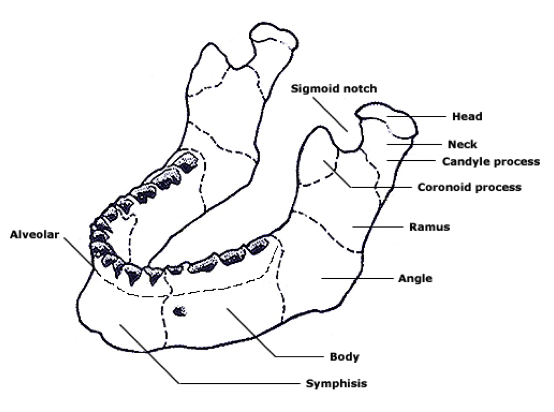
The mandible is the largest bone in the human skull, forming the lower jawline and shaping the contour of the inferior third of the face (see Image. Mandible Anatomy).[1] Articulation with the skull base at the bilateral temporomandibular joints allows a range of movements facilitated by associated muscles, including dental occlusion with the maxilla (see Image. Jaw Anatomy, Lateral View). The The TMJ is a unique joint involved in a number of important functions, including mastication and speech, 1 but more simply, it allows the articulation between the upper and lower jaws. 2 This

The Temporomandibular Joint Biology Diagrams
The temporomandibular joint (TMJ) is formed by the articulation of the mandible and the temporal bone of the cranium. It is located anteriorly to the tragus of the ear, on the lateral aspect of the face. In this article, we shall look at the anatomy of the temporomandibular joint - its articulating surfaces, ligaments and clinical correlations. The temporomandibular joint (TMJ) is a diarthrosis, better defined as a ginglymoarthrodial joint. TMJ is composed of a synovial cavity, articular cartilage and a capsule that covers the same joint. We find the synovial fluid and several ligaments. The joint is the union of the temporal bone cavity with the mandibular condyle. Anatomy
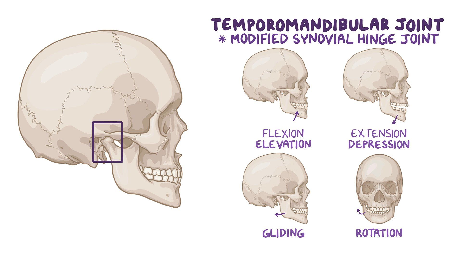
The temporomandibular joint (TMJ) is an atypical synovial joint located between the condylar process of the mandible and the mandibular fossa and articular eminence of the temporal bone. It is divided into a superior discotemporal space and inferior discomandibular space by the TMJ disc (or meniscus). In anatomy, the temporomandibular joints (TMJ) are the two joints connecting the jawbone to the skull. The lower joint compartment formed by the mandible and the articular disc is involved in rotational movement—this is the initial movement of the jaw when the mouth opens. The upper joint compartment formed by the articular disc and the The temporomandibular joint (TMJ) is a hinge type synovial joint that connects the mandible to the rest of the skull.More specifically, it is an articulation between the mandibular fossa and articular tubercle of the temporal bone, and the condylar process of the mandible.Even though the TMJ is classified as a synovial-type joint, it is atypical in that its articular surfaces are lined by
